ClearView™ 3D Dosimeter
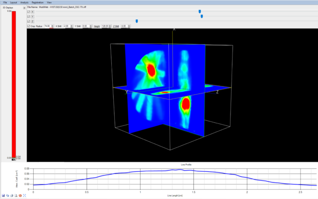
ClearView™ is a non-diffusing, radiochromic hydrogel dosimeter designed for verification of advanced radiation therapy techniques including SRS. Visualizing the intricate detail of complex dose distributions and multiple lesion dosimetry, ClearView™ aids users in confirming planned treatments are delivered accurately.
Principles of radiochromic 3D dosimetry are analogous to film (2D) dosimetry. Dose scaling with ClearView™ is achieved by using a reference dose delivery in the same dosimeter or irradiation of another dosimeter from the same manufacturing batch.
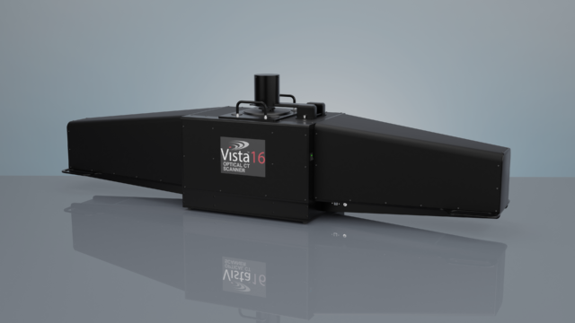
ClearView™ is intended to be used as part of an integrated 3D dosimetry system which includes an optical CT scanner such as Vista™ 16, and analysis software, Vista ACE™ (in development).
Features
Design
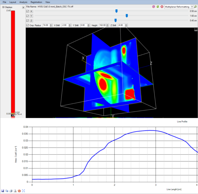
ClearView™ is optically clear, low scattering, and colourless. It contains a radiochromic indicator dye which turns purple after irradiation. The change in optical attenuation of the gel is directly proportional to the absorbed radiation dose enabling the visualization of dose distributions.
Stability
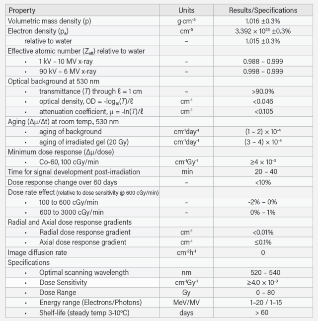
Providing utility within clinical and research settings, ClearView™ chemistry is stable for over 60 days prior to irradiation under recommended storage conditions. The signal is geometrically stable at any time after irradiation.
Linear Response
SRS applications require a high dose response dosimeter. ClearView™ dose response is linear within a range of zero to 80 Gy, creating an ideal solution for advanced radiation therapy delivery.
High Resolution
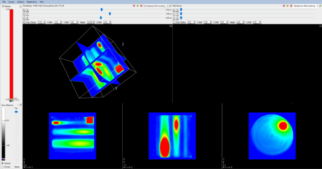
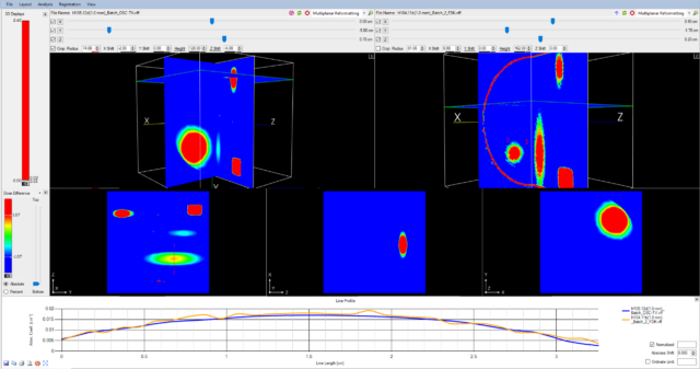
True 3D dosimetry is achievable with ClearView™. Unlike diode arrays that interpolate dose data between detectors, ClearView™ gel is geometrically accurate to a sub-micron level, giving you a high spatial resolution 3D representation of a patient’s specific treatment plan.
Additional Resources
Radiochromic Film Dosimetry with Vista 15™ Optical Cone Beam CT Scanner
BABIC, S., JORDAN, K., Radiochromic Film Dosimetry with Vista 15™ Optical Cone Beam CT Scanner, Poster presented at the 56th Canadian Organization of Medical Physicists (COMP) Annual Meeting, June 16-19, 2010, Ottawa, ON, Canada
Cone beam optical computed tomography for gel dosimetry I: scanner characterization
OLDING T., HOLMES O., and SCHREINER L. J., Cone beam optical computed tomography for gel dosimetry I: scanner characterization, Phys. Med. Biol. 55: 2819–2840, 2010
Polymer Gel Dosimetry
BALDOCK C., DE DEENE Y., DORAN S., IBBOTT G., JIRASEK A., LEPAGE M., MCAULEY K. B., OLDHAM M. and SCHREINER L. J., Polymer Gel Dosimetry, Phys. Med. Biol. 55: R1-R63, 2010
Three-dimensional Dosimetry of small megavoltage radiation fields using radiochromic gels and optical CT scanning
BABIC, S., MCNIVEN, A., BATTISTA, J. and JORDAN, K., Three-dimensional Dosimetry of small megavoltage radiation fields using radiochromic gels and optical CT scanning, Phys. Med. Biol. 54: 1-19, 2009
Three-Dimensional Dose Verification for Intensity-Modulated Radiation Therapy in the Radiological Physics Centre Head-and-Neck Phantom Using Optical Computed Tomography Scans of Ferrous Xylenol-Orange Gel Dosimeters
BABIC, S., BATTISTA, J. and JORDAN, K., Three-Dimensional Dose Verification for Intensity-Modulated Radiation Therapy in the Radiological Physics Centre Head-and-Neck Phantom Using Optical Computed Tomography Scans of Ferrous Xylenol-Orange Gel Dosimeters, Int. J. Radiation Oncology Biol. Phys., Vol. 70, No. 4, pp 1281-1291, 2008
Optical-CT gel-dosimetry II: Optical artifacts and geometrical distortion
OLDHAM, M. and KIM, L., Optical-CT gel-dosimetry II: Optical artifacts and geometrical distortion, Med. Phys. 31, 1093-1104, 2004
Optical-CT gel-dosimetry I: Basic investigations
OLDHAM, M., SIEWERDSEN, J. H., KUMAR, S., WONG, J. and JAFFRAY, D. A., Optical-CT gel-dosimetry I: Basic investigations, Med. Phys. 30, 623-634, 2003
CCD imaging for optical tomography of gel radiation dosimeters
WOLODZKO, J.G., MARSDEN, C., APPLEBY, A., CCD imaging for optical tomography of gel radiation dosimeters, Med Phys. 26: 2508-13, 1999
Optical CT reconstruction of 3D dose distributions using the ferrous-benzoic-xylenol (FBX) gel dosimeter
KELLY, B.G., JORDAN, K.J., and BATTISTA, J., Optical CT reconstruction of 3D dose distributions using the ferrous-benzoic-xylenol (FBX) gel dosimeter, Med Phys, 25:1741-50, 1998
Optical Imaging of Radiation Dose Distributions in a Ferrous-Gelatin-Xylenol Orange Gel Dosimeter
Graduate Dissertation by Richard M. Fried, Optical Imaging of Radiation Dose Distributions in a Ferrous-Gelatin-Xylenol Orange Gel Dosimeter, dissertation director Professor Alan Appleby, Rutgers University 1995
Imaging of radiation dose by visible color development in ferrous-agarose-xylenol orange gels
APPLEBY, A. and LEGHROUZ, A., Imaging of radiation dose by visible color development in ferrous-agarose-xylenol orange gels, Med Phys, 18:309-12, 1991.
