Vista™ 16 Optical CT Scanner for 3D Dosimetry
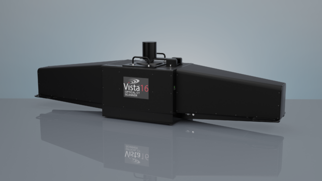
Vista™ 16 is a high-resolution optical CT scanner used to process 3D dosimeters, including radiochromic gels and solids. With the use of Clearview™ gel and Vista™ ACE processing software (in development), physicists will have a complete, true 3D dosimetry solution.
Vista™ 16 reconstructs a 3D map of optical attenuation coefficients within an object suspended in the scanner’s region of interest. This is done by capturing a series of 2D optical projections through the region of interest while the object is rotated. The 3D map is then constructed using Feldkamp-filtered back projection.
The system is designed to work with radiochromic 3D dosimeters such as ClearView™ which have an absorption peak at 590 nm. Various wavelengths are available upon request for different dosimeters and applications.
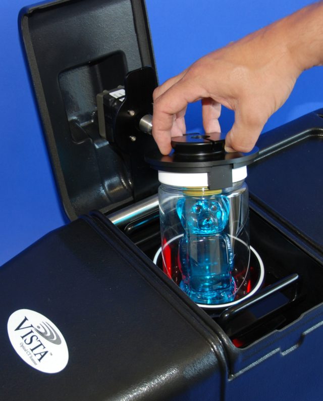
Vista™ 16 is capable of scanning objects up to 15 cm diameter by 12 cm long. Isotropic voxel sizes of 0.25 mm are achievable, providing high-resolution, true 3D dose data. VistaScan™ software is included for scanning, reconstruction and display. Data is fully accessible for treatment plan verification and research applications.
-
Specifications
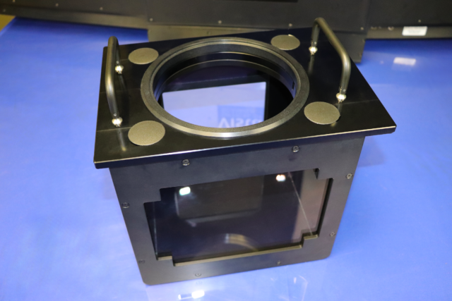
- Aquarium: Glass/Aluminum
- Region of Interest: Up to 15 cm diameter x 12 cm long
- Enclosure (WxLxH): 35 cm x 165 cm x 48 cm (including cabinet containing motor)
- Electrical – 24V DC, 2.1A ; 120 – 240 V AC medical grade switching power supply provided
-
Minimum Technical Requirements
- Operating System: Windows 7, 10
- Processor: i7, 2 GHz or better
- RAM: 4GB or more
- Graphics Card: 512 MB or better with OpenGL 2.1
- Peripheral Connectivity: USB 3.0
-
100-2016: Vista™ 16 Cone Beam Optical CT Scanner, includes:
- Scanner
- Software
- User's Guide
- Aquarium for index of refraction matching liquid
- Wavelength 530nm (other wavelengths available on request)
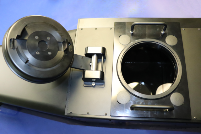
- 15 cm Rotary Stage
- 5 Polyethylene terephthalate jars (15cm diameter) Other Rotary Stage and Jar sizes available on request
-
Optional Accessories
- 500-1067: Annual Service & Software Support
- 500-1016: Aquarium (For Index of Refraction Matching)
- 500-1045: Additional Light Source (Wavelength to be confirmed prior to ordering)
- 500-1072: 10 cm Rotary Stage
- 500-1073: Dovetail Rotary Stage and Jar Clamp for Smaller Jars
- 500-1074: Jar Clamp for Smaller jars
Features
Low Stray Light
Convergent light source improves accuracy and precision of image data reconstruction.
Fast
Acquisition, reconstruction and display of optical CT images in approximately five minutes.
Workflow Efficiency
Simple on-screen controls and straightforward workflow make for an intuitive 3D dosimeter analysis solution.
Flexible
Different light sources (wavelengths) are available upon request for optimization with various dosimeters. Easily swap the liquid-filled aquarium for different index of refraction matching liquids.
Software
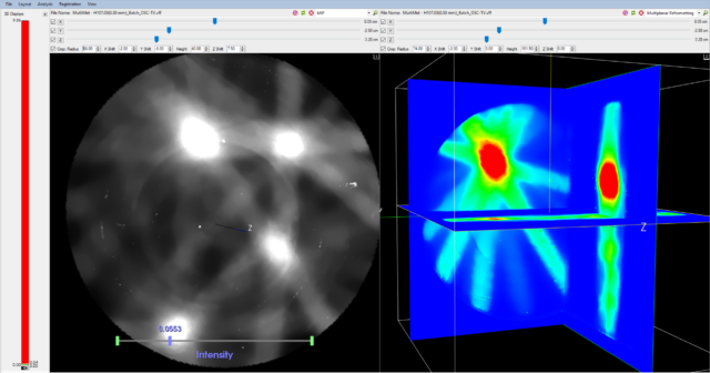
The included VistaScan™ software is used for scanner control, image reconstruction and display. Adjust scan settings such as alignment, exposure, shutter speed, and calibration parameters in real time. Data is fully accessible for treatment plan verification and research applications.
Additional Resources
Radiochromic Film Dosimetry with Vista 15™ Optical Cone Beam CT Scanner
BABIC, S., JORDAN, K., Radiochromic Film Dosimetry with Vista 15™ Optical Cone Beam CT Scanner, Poster presented at the 56th Canadian Organization of Medical Physicists (COMP) Annual Meeting, June 16-19, 2010, Ottawa, ON, Canada
Cone beam optical computed tomography for gel dosimetry I: scanner characterization
OLDING T., HOLMES O., and SCHREINER L. J., Cone beam optical computed tomography for gel dosimetry I: scanner characterization, Phys. Med. Biol. 55: 2819–2840, 2010
Polymer Gel Dosimetry
BALDOCK C., DE DEENE Y., DORAN S., IBBOTT G., JIRASEK A., LEPAGE M., MCAULEY K. B., OLDHAM M. and SCHREINER L. J., Polymer Gel Dosimetry, Phys. Med. Biol. 55: R1-R63, 2010
Three-dimensional Dosimetry of small megavoltage radiation fields using radiochromic gels and optical CT scanning
BABIC, S., MCNIVEN, A., BATTISTA, J. and JORDAN, K., Three-dimensional Dosimetry of small megavoltage radiation fields using radiochromic gels and optical CT scanning, Phys. Med. Biol. 54: 1-19, 2009
Three-Dimensional Dose Verification for Intensity-Modulated Radiation Therapy in the Radiological Physics Centre Head-and-Neck Phantom Using Optical Computed Tomography Scans of Ferrous Xylenol-Orange Gel Dosimeters
BABIC, S., BATTISTA, J. and JORDAN, K., Three-Dimensional Dose Verification for Intensity-Modulated Radiation Therapy in the Radiological Physics Centre Head-and-Neck Phantom Using Optical Computed Tomography Scans of Ferrous Xylenol-Orange Gel Dosimeters, Int. J. Radiation Oncology Biol. Phys., Vol. 70, No. 4, pp 1281-1291, 2008
Optical-CT gel-dosimetry II: Optical artifacts and geometrical distortion
OLDHAM, M. and KIM, L., Optical-CT gel-dosimetry II: Optical artifacts and geometrical distortion, Med. Phys. 31, 1093-1104, 2004
Optical-CT gel-dosimetry I: Basic investigations
OLDHAM, M., SIEWERDSEN, J. H., KUMAR, S., WONG, J. and JAFFRAY, D. A., Optical-CT gel-dosimetry I: Basic investigations, Med. Phys. 30, 623-634, 2003
CCD imaging for optical tomography of gel radiation dosimeters
WOLODZKO, J.G., MARSDEN, C., APPLEBY, A., CCD imaging for optical tomography of gel radiation dosimeters, Med Phys. 26: 2508-13, 1999
Optical CT reconstruction of 3D dose distributions using the ferrous-benzoic-xylenol (FBX) gel dosimeter
KELLY, B.G., JORDAN, K.J., and BATTISTA, J., Optical CT reconstruction of 3D dose distributions using the ferrous-benzoic-xylenol (FBX) gel dosimeter, Med Phys, 25:1741-50, 1998
Optical Imaging of Radiation Dose Distributions in a Ferrous-Gelatin-Xylenol Orange Gel Dosimeter
Graduate Dissertation by Richard M. Fried, Optical Imaging of Radiation Dose Distributions in a Ferrous-Gelatin-Xylenol Orange Gel Dosimeter, dissertation director Professor Alan Appleby, Rutgers University 1995
Imaging of radiation dose by visible color development in ferrous-agarose-xylenol orange gels
APPLEBY, A. and LEGHROUZ, A., Imaging of radiation dose by visible color development in ferrous-agarose-xylenol orange gels, Med Phys, 18:309-12, 1991.
